|
Cervical Disease and Neoplasia |
|
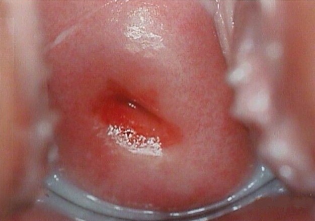
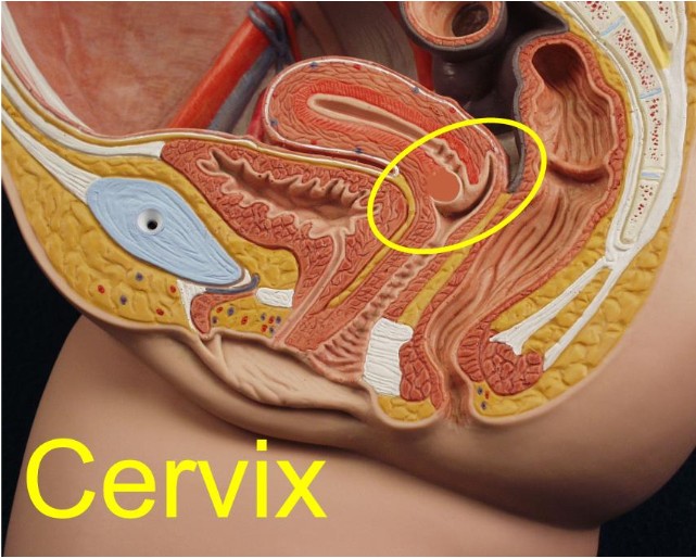
The opening of the uterus is called the cervix. While the cervix is considered a portion of the uterus, it is functionally and histologically quite different. It is composed of dense connective tissue, with very little smooth muscle. The body of the uterus, in contrast, is primarily smooth muscle. The cervix is located at the top of the vagina and is easily visualized by inserting a vaginal speculum fully into the vagina and opening the blades. The firm, smooth, pink structure appearing at the end of the vagina is the cervix. The cervix is of clinical significance because of:
|
||||||||||||||||||
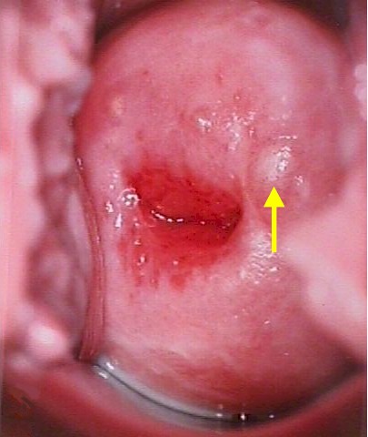 Nabothian Cyst |
Nabothian Cysts Two normal anatomic variations are responsible for considerable clinical concern among the less experienced who examine women, Nabothian cysts and cervical ectropion. Nabothian cysts from when the secretions from functional glandular epithelium become trapped below the surface of the skin. This can occur because of the normal deep infolding of the endocervical epithelium. It also may occur when the squamous exocervical epithelium covers over the mucous-producing endocervical epithelium (squamous metaplasia). It is seen more commonly after childbirth and sometimes occurs concomitantly with cervicitis. Clinically, Nabothian cysts are seen or felt as firm nodules on the cervix. If close to the surface, they care clearly seen as cystic. If the diagnosis is in doubt, puncturing them will release clear, mucoid material. It is not necessary to do this, however, as the diagnosis is rarely in doubt. Sometimes, particularly if large, Nabothian cysts will be seen to have a significant blood vessel or two coursing over the surface. These are of no concern, though if tampered with, they may bleed. |
||||||||||||||||||
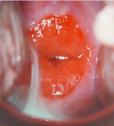 Cervical Ectropion |
Cervical Ectropion Inexperienced examiners are sometimes frightened to see a large, red, somewhat friable lesion occupying the central portion of the cervix and surrounding the cervix. This is not a lesion...it is cervical ectropion. The red color is from the shallow, vascular, mucous-producing endocervical epithelium which has grown out onto the face (exterior portion) of the cervix. Over time, the ectropion may enlarge or diminish in size, as the squamocolumnar junction changes its relative position on the cervix. With pregnancy, the cervix tends to evert, making the ectropion larger. In menopause, the SQJ tends to recede back up the cervical canal, making the ectropion get progressively smaller before disappearing completely. The fact that a cervical ectropion is present is of no clinical concern. It needn't be treated and can safely be ignored. If the ectropion is causing symptoms (eg., post-coital bleeding), or in the presence of recurrent cervical infections (cervicitis), then the ectropion can be treated by any means that safely eliminates the most superficial layer of cells, facilitating the inward growth of the surrounding squamous mucosa. Among these treatments are cryosurgery, chemical cautery (AgNO3), electrocautery, thermal cautery, LEEP, and laser ablation. |
||||||||||||||||||
 |
Cervicitis Cervicitis means an infection of the cervix. This usually involves the glandular elements (columnar epithelium) of the cervix and is usually caused by organisms found in the vagina. Sometimes it is caused by such sexually transmitted diseases as chlamydia or gonorrhea. Most cases of cervicitis are unnoticed by the patient, but some notice painful intercourse, persistent vaginal discharge, aching pelvic pain or menstrual cramps. The diagnosis can be confirmed by palpation of the cervix. Normally, this doesn't hurt, but in the case of cervicitis, compression or movement of the cervix causes some discomfort. When clinically warranted, testing for STDs can be helpful before initiating treatment. If found, they should be individually treated. A simple course of oral antibiotics can be effective at resolving the cervicitis, although if there is extensive cervical ectropion, the cervicitis may return. For recurrent cervicitis, some form of cervical ablation is usually needed, in addition to a short course of antibiotics, to permanently resolve the problem. |
||||||||||||||||||
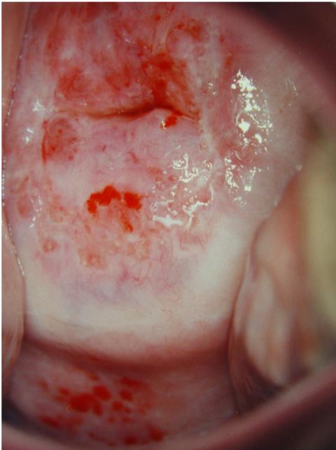
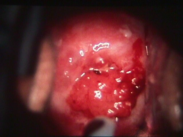 Invasive Squamous Cell Cancer |
 Cervical Cancer Cervical CancerInvasive cancer of the cervix is a relatively uncommon malignancy among women, representing about 2% of all new cancers. This is in contrast to the more common female cancers (breast 30%, lung 13%, colon 11%). It is less common than uterine (6%). ovarian (4%), and bladder (3%) cancer. Pap smear screening has had a dramatic impact on the incidence of cervical cancer. Initially throught to promote early diagnosis of cervical cancer, Pap screening has been most helpful in detecting the pre-malignant changes that, when treated, are effective in preventing the actual development of cervical cancer. Most cases of invasive cervical cancer occur among women who have not been screened with Pap smears or who have not had a Pap smear in many years. Those at increased risk for cervical neoplasia include women with multiple sexual partners, HPV infection (particularly the high risk HPV types), cigarette smokers and those with impaired immune systems. The most common symptom of cervical cancer is abnormal vaginal bleeding, either spontaneous or provoked by intercourse or vigorous physical activity. In advanced cases, some patient will notice back or flank pain provoked by ureteral obstruction and hydronephrosis. The diagnosis is usually confirmed by biopsy, but is suspected if a friable, visible, exophytic lesion is seen on the cervix. Endophytic lesions tend to invade deeply into the cervical stroma, creating an enlarged, firm, barrel-shaped cervix. Initial metastases are to the parametrial tissues and lymph nodes. Later, in addition to aggressive local spread into the upper vagina, rectum and bladder, hematogenous spread to the liver, lungs, and bone occurs. Following diagnosis of cervical cancer, staging of the cancer is performed to assist in selecting the best treatment. Stages (0-IV) are defined by the International Federation of Gynecology and Obstetrics, and include:
In addition, there are subcategories within each stage. Treatment can consist of surgery, radiation therapy, and sometimes adjuvant therapy. The best option for treatment depends on the stage of the cancer. Surgery seems to work best on Stages I and IIA (no obvious parametrial involvement). Radiotherapy seems to work better for more advanced stages. Complications of either approach include bladder and bowel fistula formation, and loss of vaginal length. |