|
Introduction to Ultrasound |
 Humans can hear sound with frequencies of 20 to 20,000
cycles per second (20-20,000 Hertz or Hz). Any frequency higher than
that is called ultrasound.
Humans can hear sound with frequencies of 20 to 20,000
cycles per second (20-20,000 Hertz or Hz). Any frequency higher than
that is called ultrasound.
Ultrasound is diagnostically useful in medicine two modalities, continuous energy and pulsed energy:
-
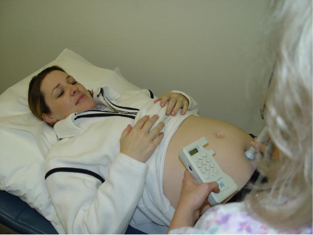
Continuous sound energy uses a steady sound source, and has applications that include fetal heart beat detectors and monitors. This Doppler ultrasound can also be used to evaluate blood flow through different structures.
-
Pulsed sound energy utilizes a quick blip of sound (like a hand clap), followed by a relatively long pause, during which time an echo has a chance to bounce off the target and return to the transducer. Through electronic processing of the returning sounds, a two-dimensional image can be created that provides information about the tissues and objects within the tissues.
 Physics
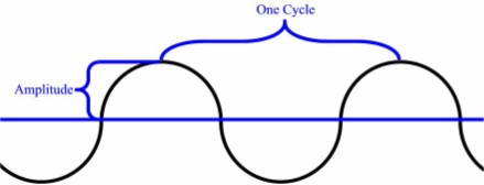
Ultrasound is a form of mechanical energy that in many respects behaves
according to the properties of wave-form physics. For this reason,
terminology of wave-form physics is usually applied, including such
terms as wave amplitude and cycle frequency.
Remember that sonic energy is not identical to electromagnetic radiation and that while they share some of the same properties, sound can behave differently, particularly at extreme ends of the spectrum, when passing through complex media, or when interfered with by conflicting sounds.
Doppler Ultrasound
The Doppler Principle is easiest illustrated by listening to a train
approaching. As it gets closer, you hear the horn at a certain pitch
(frequency). As the train passes, you hear the sound of the horn drop to
a lower pitch. You have just experienced the Doppler Principle.
Consider an object that generates a sound. At rest, the sound frequency is constant. If the object moves towards you, the sound that you hear will seem a little higher in frequency. If the object moves away from you, the sound will have a lower frequency.
Fetal heart beat detectors generate a constant sound. Some of the sound is reflected back toward the transducer. The frequency of the outgoing sound and incoming sound is the same, UNLESS the object is moving (either toward the transducer or away from it. Blood passing through the heart or major placental vessels will reflect back sound that is a slightly different frequency (higher or lower) than the frequency generated by the machine. Because they are of a slightly different frequency, they never line up evenly, except every now and then when the both incoming and outgoing sound energies line up perfectly. The "beat frequency" happens to be in the audible range (less than 10,000 Hz), and can be detected and amplified.
So when you are listening to a fetal heart beat, you are not actually hearing the sound of the heart. You are hearing the beat frequency generated by the interference between the outgoing ultrasound frequency and the incoming ultrasound frequency, that are slightly different because of the movement of the heart wall and blood flowing through the heart and large vessels.
 Clap-Echo System |
Pulsed Ultrasound
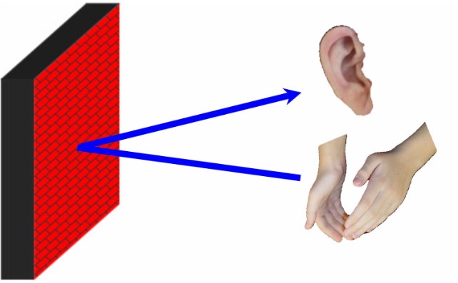
If you clap your hands in a large, empty room, you may hear the echo
from the sound of the clap bouncing off the far wall and returning to
you. Pulsed ultrasound imaging technology is similar to the clap and
echo.
If you could accurately measure the time it took from your handclap to the time you heard the returning echo, you could calculate how far the sound has traveled, and by inference, how far away the wall is from you.
distance = (time) x (speed of sound in air)
Of course, you have to remember that the distance traveled by the sound is twice the distance to the wall...the sound had to travel out to the wall and then back to you (a round trip).
 A-Mode Ultrasound |
A-Mode Ultrasound
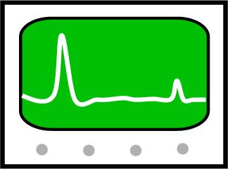
On an oscilloscope, this simple clap-echo system would look like this.
The initial spike from the clap would be followed some time later by the echo. The earliest "A-Mode" ultrasound machines worked in this way. You could know how far the echo has traveled, and how loud the echo was when it got back to you.
 There were (and are) several problems with this simple
system:
There were (and are) several problems with this simple
system:
-
You don't know the exact direction it came from.
-
You don't know for sure what the echo bounced off of.
-
You don't know what the object generating the echo looks like.
 B-Mode Ultrasound
Imaging
B-Mode Ultrasound
Imaging
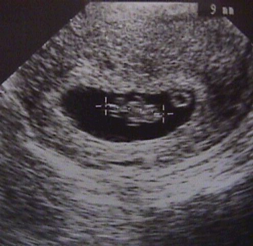
B-Mode ultrasound imaging collects the same information, but adds a
sense of direction (where the echo is coming from in a two-dimensional
plane) as well as the memory to recall all the different echoes, strong
and weak.
This image becomes recognizable, particularly with practice. The recognizable image can then be evaluated for abnormalities, and measured.
B-mode imaging was the first practical application of ultrasound for diagnostic purposed.
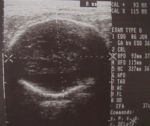
Real Time Imaging
The ability to appreciate the structures within a two-dimensional image
is very much enhanced by visualizing the changes that occur within that
image over time.
A real-time image is still a 2-dimensional view, but one that is constantly updated. This then becomes 3-dimensional imaging (height, width, and time).
Real-time sonography is most useful when the visualized object is moving (like a fetal heart), but is also valuable when the transducer beam is swept through the object, enhancing the operator's appreciation for details, texture, and shape.