|
Hysterosalpingogram |
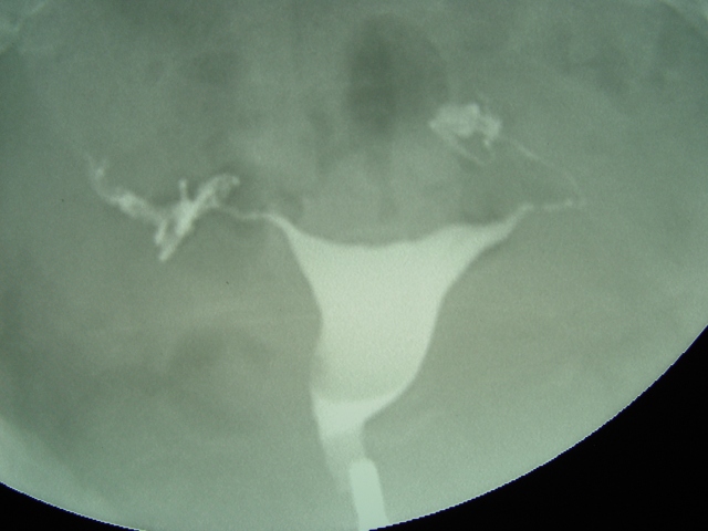
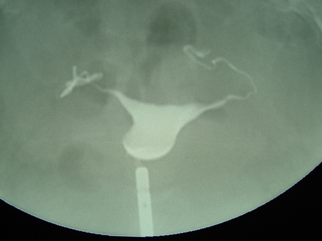 1. A metal catheter is passed through the cervix and dye has filled the uterine cavity. It is just starting to fill the tubes.
|
The interior of the uterus and fallopian tubes can be evaluated with an
x-ray dye study call a hysterosalpingogram. This is often performed as
part of an infertility evaluation. It's purpose is to identify anatomic
abnormalities (submucous fibroids, endometrial polyps, uterine
malformations, blocked fallopian tubes and others). It is performed during the early proliferative phase (after cessation of menses but before ovulation) to avoid disrupting an early pregnancy. Radio-opaque dye is slowly injected through the cervix and into the uterine cavity under a small amount of pressure. The dye is followed with flouroscopy as it fills the uterine cavity and then travels retrograde into the fallopian tubes. The internal diameter of the tube is identified and ultimately dye spills out the finbriated end and into the abdominal cavity. |