|
Wet Mount |
|
|
While it is often possible to correctly guess the cause of a vaginal discharge,
based on history and/or physical exam, it is sometimes useful to use laboratory
skills to confirm a clinical impression.
A wet mount is the suspension of a small amount of vaginal discharge in a liquid medium. Two liquids are commonly used, normal saline and potassium hydroxide. Each has it's own unique properties that make it useful in this setting:
|
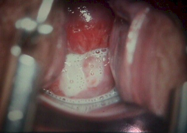
 Obtain a sample of the vaginal discharge. |
Obtain a Specimen I prefer to use a non-wooden collector because I avoid the tiny wood fragments that contaminate the microscopic field. The discharge need not be processed immediately, but can wait until you have completed the rest of your exam. You can't wait indefinitely, though. If the discharge dries completely before you can process it, the information you obtain will be of less value. |
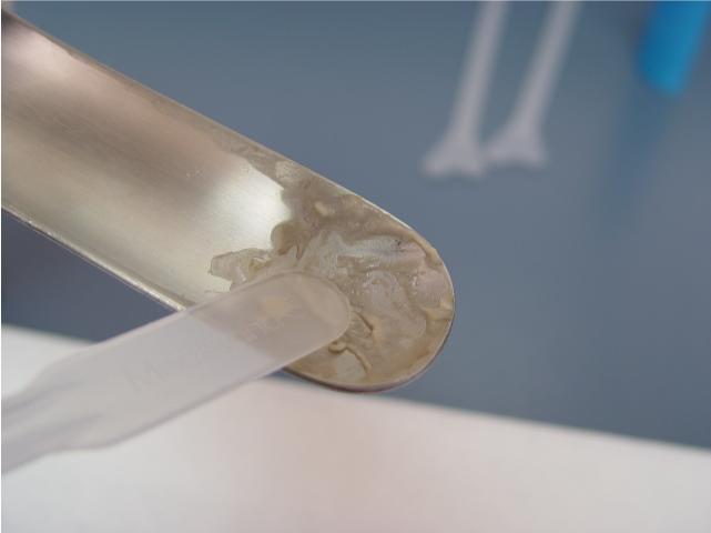
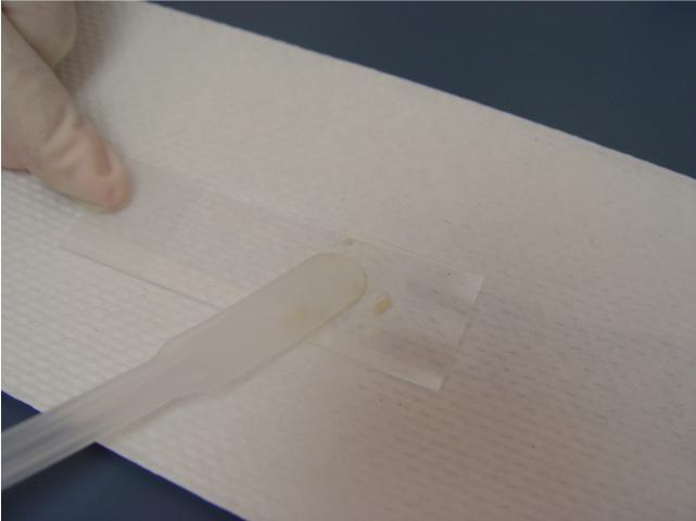 Place a small amount of the discharge on a glass slide. |
Put a Tiny Amount of Discharge on a
Microscope Slide Later, when you view it under the microscope, it will be spread as thin as a single cell. If you start off with too much discharge, it will make it harder for you to see the individual structures you need to evaluate. Since you will actually be making two wet mounts (one of normal saline and one of KOH), you can put some discharge on each of two slides. Others prefer to use just a single slide, and they put a bit of discharge at each end of the glass slide, to keep them separate. In this case, it is important to keep them far enough apart that when you add the solutions later, there will be no mixing of the NaCl and KOH. |
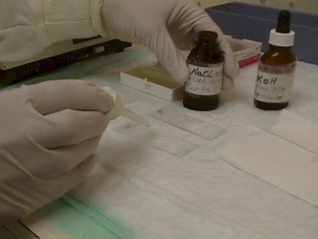 Add a drop of normal saline and add a coverslip. Prepare a second, similar slide using potassium hydroxide. |
Add NaCl Add one drop of Normal Saline (0.9 percent NaCl) to the drop of discharge. Mix well on the slide. This is the slide you will use for identifying Trichomonas and bacterial vaginosis (BV). The drop of NaCl should fall freely to the slide. Avoid touching the dropper to the slide, or touching the drop to the slide before the drop is released from the dropper. Either of these can result in your contaminating the NaCl bottle with material from the discharge. Prepare a second slide in the same way, using 10 percent Potassium Hydroxide (KOH). This is the slide you will use to identify yeast. As you are mixing the KOH with the discharge, you may notice an unpleasant amine smell. This is called a positive "whiff test" (you caught a whiff of bad smell), and indicates the presence of bacterial vaginosis. |
 The KOH dissolves the cell membranes faster if the slide is heated. |
Add Coverslips Place glass coverslips over the glass slides. Remove any excess fluid with tissue paper. In order for the KOH to be effective in dissolving the cell membranes of everything except yeast, you need to allow some time. A minute or two may be enough. If you are in a hurry, you can speed the process by heating the KOH slide with a match or lighter. The elevated temperatures will speed the dissolving process and the glass slide cools quickly enough that you can place it under the microscope as soon as you've finished heating it. Do not warm the saline slide as you will stop flagella movement and coagulate the proteins of the structures you are trying to identify. |
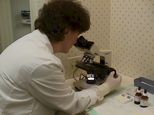 |
Microscopic Evaluation Examine the prepared slides under a microscope. Experienced practitioners often find the lowest power (about 40X) works the best. Others will start at low power and then move to slightly higher power (about 100X). The magnification is determined by multiplying the power of the eyepiece (typically 10X) by the power of the objective lens (4X, 10X, 40X, 80X) to get the various possible total magnifications (40X, 100X, 400X, and 800X in this example.) |
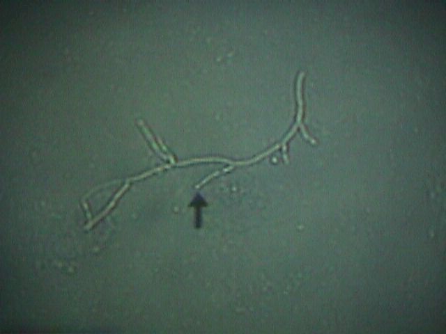
 Yeast on low power
|
Yeast Yeast (Candida, Monilia) is best identified with the KOH slide. After the cell membranes are dissolved, the typical branching and budding yeast cells can be seen. Sometimes, it has the appearance of a tangled web of threads. At other times, only small branches will be seen. Yeast are normal inhabitants of the vagina, but only in very small numbers. If you visualize any yeast in your sample, it is considered significant.
|
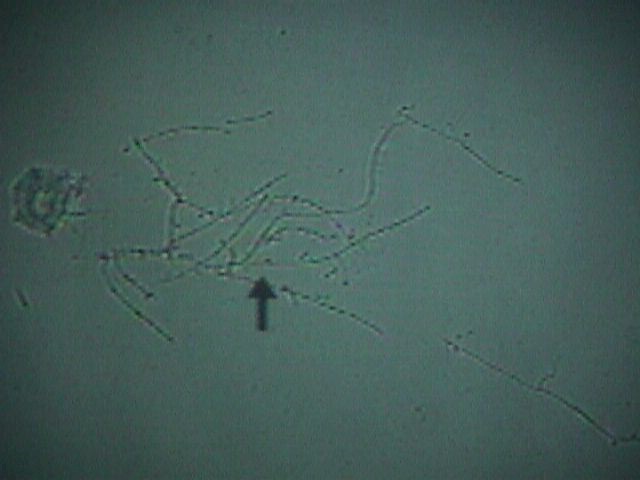
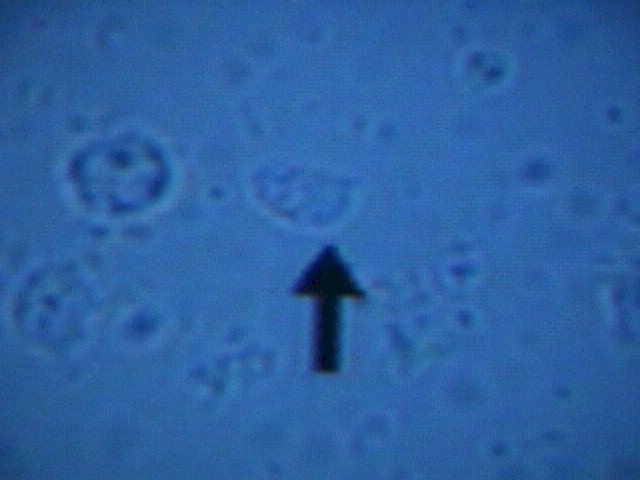 Trichomonad (arrow) next to a white blood cell (to the left) |
Trichomonas Trichomonas is best seen on the Normal Saline slide. These protozoans are about the same size as a white blood cell (a little smaller than a vaginal epithelial cell), but their violent motion is striking and unmistakable. |
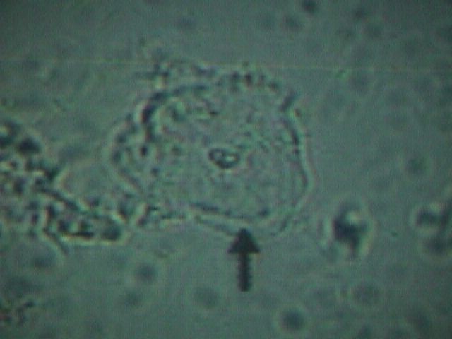
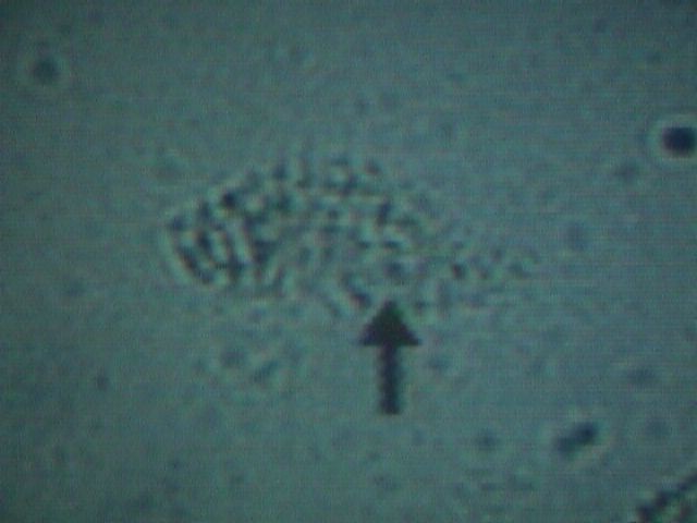 Clue Cell showing bacteria studding the surface of this vaginal epithelial cell.
|
Bacterial Vaginosis Bacterial vaginosis (also known as Gardnerella, hemophilus, or non-specific vaginitis) is characterized by the presence of "clue cells" visible at both low and medium power. These clue cells are vaginal epithelial cells studded with bacteria. It resembles a pancake that has fallen into a bowl of poppy seeds, but on a microscopic level. A normal vaginal epithelial cell is clear, with recognizable contents, and sharp, distinct cell borders. A clue cell appears smudged, with indistinct contents and fuzzy, poorly defined borders. |