|
Vulvar Biopsy |
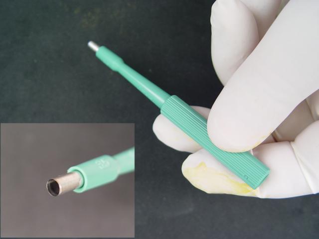 |
A vulvar biopsy is an excellent method of obtaining microscopic information whenever there is a vulvar lesion of uncertain significance, or in the presence of persistent symptoms, such as vulvar irritation or itching. There are many good ways to perform a vulvar biopsy. Among them is the use of the Keyes Punch, demonstrated here. The Keyes Punch has a sharpened, round tip, designed to take a vertical core from the epithelium.
|
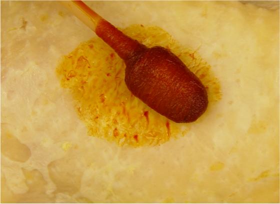 |
After selecting the biopsy site, repare the skin with antiseptic, such
as alcohol, iodophore, or other suitable material. Take care in selecting the biopsy site. Vulvoscopy (using the colposcope to look closely at the vulva) can help in choosing the site to biopsy. Application of acetic acid helps here, as it does on the cervix, although it takes considerably longer for acetowhite lesions to appear. Another technique is to pain the vulva in toluidine blue dye, followed by an acetic acid rinse. Areas of increased metabolic activity (increased DNA density) will hold the blue stain more than the surrounding normal tissue. Areas to avoid:
|
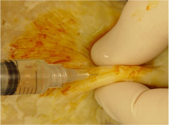 |
Pinch the skin between your thumb and forefinger. This will bunch up the skin and stabilize it. Then inject a small amount of local anesthetic. In this example, I am using about 2 cc of 1% lidocaine. Some prefer to add epinephrine, but I don't think it matters either way. |
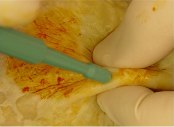 |
While maintaining your hold on the skin, direct the Keyes Punch straight
down into the skin. Rotate the Keyes Punch, rolling it clockwise, then
counterclockwise, to assist it in penetrating through the epithelium. This action will produce a nice core-shaped specimen that is very small, but descends through the full thickness of the epithelium and into the subcutaneous tissues below. |
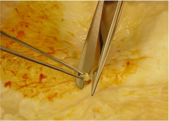 |
Grasp the tissue plug with forceps and elevate it slightly above the skin. Usually, it will still be connected at the base. Use scissors to cut across the base, freeing the plug from the subcutaneous tissues. |
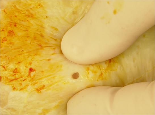 |
You will be left with a tiny, circular hole through the epithelium. When it heals completely, the normal contraction process will result in the tiniest dot of a scar, not generally visible without magnification and knowing where the biopsy site is located. |
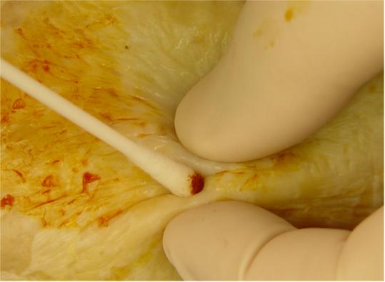 |
Usually, the base of the epithelial defect bleeds some. You can often
control this with direct pressure for a minute or two, but I generally
apply either Monsel's solution (plus direct pressure) or touch a silver
nitrate stick to the base of the defect (and then apply direct
pressure). I don't remember ever having to place a suture to control the bleeding, and don't recall any patient returning because the bleeding, once controlled, started up again. |