Inspect
Raise Arms
Tighten Pectoralis Muscles
Palpate
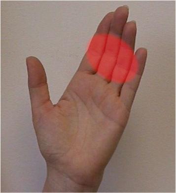
Hand

Use a Circular Motion

Sometimes, Use Two Hands
Strip the ducts, looking for discharge
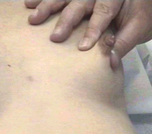
Check Axilla
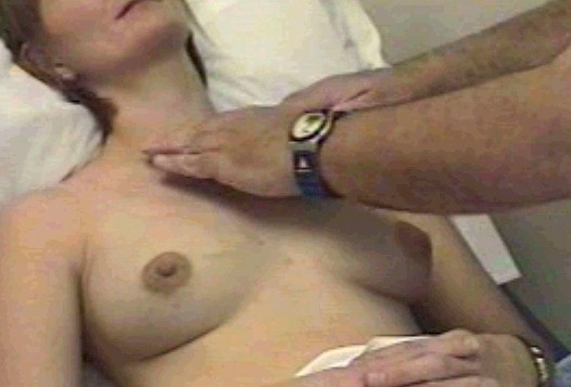
Supraclavicular Area

Reclining Palpation

Reclining Axilla Check
Reclining Supraclavicular Exam
Check for Discharge |
|

Breast examination A breast examination consists of inspection and palpation of the breasts
to identify abnormalities. Some breast examinations are focused on
specific issues while others are more general. Although there are many
good ways to examine the breasts, one of them will be presented here.
 The breasts may be examined while the patient is sitting or reclining.
Sometimes it may be desirable to do both. The breasts may be examined while the patient is sitting or reclining.
Sometimes it may be desirable to do both.
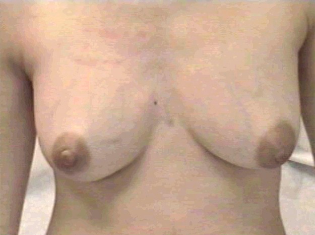
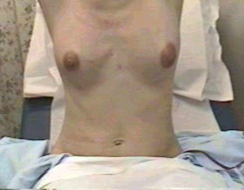
With the patient in a sitting position, inspect the breasts visually.
While generally symmetrical, most breasts are slightly asymmetrical in
respect to size, shape, orientation, and position on the chest wall.
Inspect for:
- Visible masses (change in contour)
- Skin dimpling
- Nipple retraction
- Redness
Have her raise her arms while you continue to watch the breasts.
- An underlying malignancy can fix the skin in place.
- Raising the arms will accentuate these changes.
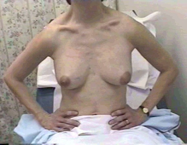
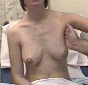
Have her flex the pectoralis major muscles. A simple way to do this
is have her place her hands on her hips and squeeze inward. Another way
is have her place her palms together (praying position) and squeeze the
palms together.
- With flexion of the underlying muscle, areas of breast tissue that
are fixed in place will move with the muscle, while the rest of the
breast will not.
- Suspicious areas will appear as a dimpling of the skin while she
flexes these muscles.
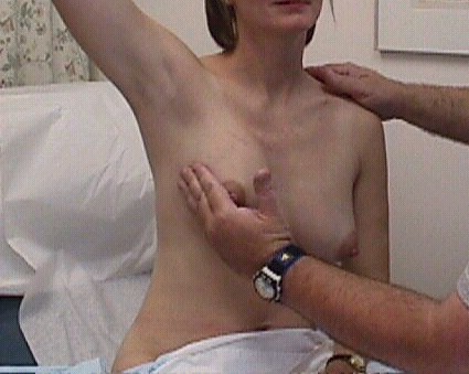
Ask her to raise her arm over her head.
- This has the effect of stretching and tightening the pectoralis
major muscle that lies directly undeneath the breast.
- Functionally, this places the breast on a fairly solid, fairly
flat surface, making it easier to palpate abnormalities.
With the patient's arm raised over her head, palpate for lumps,
masses or thickenings.
- Breast tissue is normally somewhat nodular or "lumpy,"
particularly in the upper outer quadrant.
- You are looking for a dominant mass.
- Some have suggested that you are looking for "a marble in a bag of
rice."
Palpate the breast using the proximal and middle phalanges of the
fingers.
- The palm of the hand is too insensitive to detect subtle changes
in breast texture.
- The fingertips are too sensitive and will focus on the normal
granularity of the breast tissue rather than the more worrisome
masses.
Move your hand in a circular motion while pressing into the breast
substance.
- Making these small circles will help you identify mass occupying
lesions.
- Cover the entire breast in a systematic fashion, including the
tail of the breast that extends up into the axilla.
With smaller breasts, palpation with one hand will give good results.
- When breasts are larger or pendulous, it may be useful to use two
hands, compressing the breast tissue between them.
Some examiners have the woman raise both arms above the head during
the examination. Others have her raise only one arm, leaving the other
arm down.
- Many patients feel exposed a vulnerable during this examination.
It is not a comfortable feeling for them. They may feel more
comfortable if only one arm is raised as they will feel less
vulnerable.
- Similarly, leaving one breast covered while you examine the other
breast will often make the patient feel more comfortable during the
exam. In cases where you are going back and forth, comparing findings
from one side to the other, one-sided draping may not be practical.
Check the axilla for masses or palpable lymph nodes.
- It is relatively common to find palpable lymph nodes in the axilla
of normal patients.
- These most often are the result of trauma to the arm, such as
small cuts or scrapes
- They usually disappear by themselves over a 1-2 month period of
time.
- Palpable axillary lymph nodes that are present because of an
underlying malignancy will not gradually regress over time.
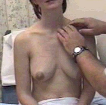
Check the supraclavicular area for palpable masses.
- The supraclavicular area can be an area of spread of breast
malignancy.
- Palpable masses or lymph nodes in this area can be a sign of
underlying malignancy.
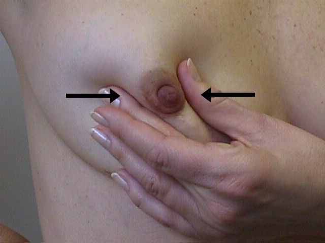
Stripping the ducts toward the nipple will cause any secretions to be
expressed.
- This should be done firmly, but not so hard as to cause discomfort
or pinching.
- With effort, you will almost always be able to bring a drop or two
of breast secretions to the surface. This is normal and the secretions
will be clear, milky, or have a slight greenish tinge.
- Bloody discharge is always considered a danger sign.
- Large amounts (many drops) of secretions are not considered normal
and usually require further investigation.
After completing the examination on one side, move to the other
breast and repeat the examination.
- Experienced examiners will frequently go back to the first breast
to compare findings from one side to the other.
- A thickening that is symmetrically present in both breasts is
usually of no significance.
- A thickening that is present in only one breast is more worrisome.
The breast examination can also be performed with the patient
reclining.
- The patient may be flat, or semi-reclining with the head and trunk
partially raised.
Go through the same systematic examination described above.
Sometimes, lumps or masses are better appreciated in the reclining or
semi-reclining position.
|