|
Fluid-Enhanced Ultrasound |
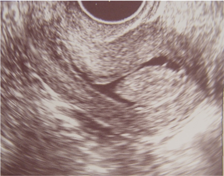 Endometrial Polyp |
One technique that is particularly useful in the office evaluation of abnormal uterine bleeding is sonohysterography, or fluid-enhanced ultrasound. A thin catheter is introduced through the cervix and into the uterine cavity. Then, transvaginal ultrasound scanning is performed while sterile saline is injected through the catheter into the cavity. This separates the uterine walls and outlines any intrauterine masses, such as polyps or fibroids. The uterine lining can also be carefully evaluated for thickening or projections. Once identified, these abnormalities can be removed through D&C. Conversely, those whose cavities and lining are normal will not usually not benefit from D&C or hysteroscopy as no abnormality will be found. |