|
Prenatal Care |
|
|

First Prenatal Visit
At the first prenatal visit, take a careful history, looking for factors
that might increase the risk for the pregnant woman.
Many providers use a questionnaire, filled out by the patient, as a starting point for this evaluation. A sample Prenatal Registration and Obstetrical Questionnaire form can be used for this purpose.
One important aspect of prenatal care is education of the pregnant woman about her pregnancy, danger signs, things she should do and things she should not do.
Many providers find it useful to give the woman printed material covering these issues that she can take with her. This allows her to read the material at a later time and to refer to it whenever she has questions. A sample Prenatal Information form can be printed and used.
Early in pregnancy, often at the first prenatal visit, a complete physical exam is performed. At that time, a Pap smear and cervical cultures are obtained. In many practices, an ultrasound scan is done at or shortly after the first visit to:
- Confirm intrauterine pregnancy placement
- Confirm fetal viability
- Confirm the number of fetuses
- Provide a highly reliable estimate of gestational age
It is valuable to document your findings in a structured flow-sheet. Many offices and hospitals have developed their own, but one is shown here:
There are so many issues to cover during the first
prenatal visit (history, physical, labs, patient education,
paperwork), that many physicians schedule two "first prenatal visits."
EDC
Based on the history, physical exam and ultrasound scan (if done), it is
important to establish a gestational age and estimated date of
confinement (EDC, or "Due Date").
You may use the last menstrual period, if known, reliable, and the patient has a history of regular periods. Add 280 days (40 0/7 weeks) to the LMP and this will give you her EDC. This assumes that she ovulated on day #14 of her last menstrual cycle. To assist you in making this calculation, I'm enclosing a LMP to EDC conversion chart here:
You may take the LMP, add 7 days and subtract 3 months. This is a rough but usable adaptation of the 280 day rule. It has the same limitations.
You may measure the fundal height (distance from the symphysis to the top of the uterus). That distance in centimeters is roughly equal to the weeks gestation of the patient.
Estimates of gestational age and EDC are best done early in pregnancy when the patient's memory is the best, and the variation is uterine size and fetal size is small.
Initial Lab Tests
Shortly after registration, initial laboratory tests are ordered.
Later in pregnancy, other tests are usually performed. Physician
preference and patient population guide some of the choice of these
tests, but commonly-ordered tests include:
- Hemoglobin and hematocrit (HGB/HCT)
- White blood cell count (WBC)
- Urinalysis (UA)
- Blood type and Rh
- Hepatitis B Screen
- Rubella Titer
- Atypical antibody screen
- Thyroid Stimulating Hormone (TSH)
- Serologic test for syphilis (RPR or VDRL)
- HIV
- Gonorrhea
- Chlamydia
- Pap
- Other lab tests as indicated by individual circumstances. For example, Sickle screening for black patients, Tay-Sachs screening for Ashkenazi Jewish patients, and thalassemia screening for patient's of Mediterranean extraction.
Subsequent Lab Tests
- Serum AFP at 15-18 weeks
- Targeted (Level II) ultrasound scan for women at high risk at 16-20 weeks
- Hbg/Hct at about 28 weeks
- Glucose screening at about 28 weeks (50 g oral load with 1-hour glucose test)
- Antibody screen and Rhogam for Rh negative women at 28 weeks
- Vaginal/rectal culture for Group B Strep at about 36 weeks
- every 4 weeks until 28 weeks' gestation
- every 2-3 weeks until 36 weeks' gestation
- every week from 36 weeks to delivery
At these visits, you will want to ask the patient about any interval changes. You'll also want to know about any vaginal discharge or bleeding, fetal movements, and uterine contractions.
At each visit, perform a limited physical exam, consisting of weight, blood pressure, edema, fundal height, fetal heart rate, and note the presence or absence of proteinuria and glucosuria. At times, it may be important to determine fetal orientation.
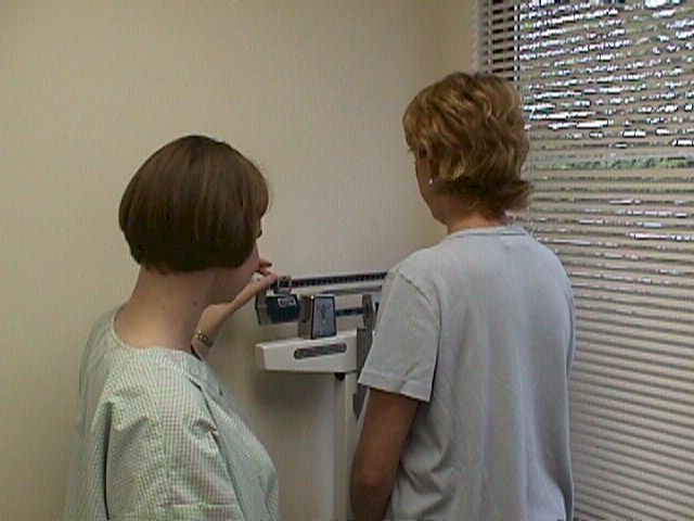 Check weight
Typical weight gain is about a pound a week. This means 30 to
40 pounds for the entire pregnancy, although some physicians feel the
ideal weight gain should be closer to 25 pounds.
Weight gain is usually slow during the first 20 weeks. Then, there is usually rapid weight gain from 20 to 32 weeks. After that, weight gain generally slows and there may be little, if any weight gain during the last few weeks.
Too little weight gain (below 13 pounds) leads to concerns that the baby may not be getting enough nutrition.
Too much weight gain leads to concerns about soft tissue distocia during labor and difficulty with restoring normal weight after delivery.
If there is sudden weight gain (more than 2 pounds in a week or more than 6 pounds in a month), this may be associated with the development of fluid retention due to pre-eclampsia (toxemia of pregnancy).
Blood Pressure
Measure the blood pressure at each prenatal visit. Significant
cardiovascular changes occur during pregnancy, including a 50% increase
in blood volume, 50% increase in cardiac output, significant reduction
in peripheral resistance, and a mild, sustained tachycardia. While these
changes are taking place, I would make the following generalizations
about blood pressure:
-
Blood pressure in early pregnancy will usually reflect pre-pregnancy levels.
-
During the 2nd trimester, maternal blood pressures usually fall below prepregnancy levels.
-
During the 3rd trimester, blood pressure usually goes back up to the pre-pregnancy level.
-
Any sustained BP of 140/90 or greater is considered significant and may indicate the development of pre-eclampsia.
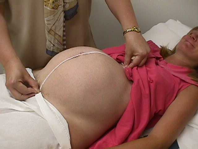 Fundal Height
Use a tape measure to record the size of the uterus. The fundal
height, measured in cm, should be approximately equal to the weeks
gestation, from mid-pregnancy until near term (MacDonald's Rule).
Measurements falling within 1-3 cm of the expected value are considered
normal. Fundal heights 4 cm different than expected are considered
abnormal and suggest the need for further investigation.
If the measurements are too small, consider:
- Your estimate of gestational age may be incorrect
- There may be very little amniotic fluid (oligohydramnios).
- The baby may be small for gestational age (or growth retarded)
- The baby may be normal, but simply constitutionally small.
If the measurements are too big, consider:
- Your estimate of gestational age may be incorrect
- There may be too much amniotic fluid (polyhydramnios)
- The baby may be large for gestational age (as is seen in gestational diabetes)
- The baby may be normal, but constitutionally large.
|
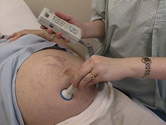
Listen for the heartbeat
The normal rate is generally considered to be between 120 and
160 beats per minute.
-
The rates are typically higher (140-160) in early pregnancy, and lower (120-140) toward the end of pregnancy.
-
Past term, some normal fetal heart rates fall to 110 BPM.
-
There is no correlation between heart rate and the gender of the fetus.
Use a coupling agent (eg, Ultrasound jel, surgical lubricant, or even water) to make a good acoustical connection between the transducer and the skin.
Doppler fetal heartbeat detectors are moderately directional, so unless you happen to aim it directly at the fetal heart initially, you will need to move it or angle it to find the heartbeat.
Confirm a normal rate, and listen for any abnormalities in the rhythm of the fetal heart beat.
Check for edema
Swelling of the feet, ankles and hands is common during
pregnancy. If mild, and in the absence of hypertension, the patient can
be reassured that:
-
This is a normal occurrence
-
While unpleasant, it is not dangerous
-
It will resolve spontaneously after the baby is born.
-
It may take weeks for the edema to resolve after delivery.
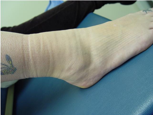 Edema of the ankle and foot, with marks from the elastic of the patient's socks indenting the skin. |
Facial edema, severe pedal edema, or any sudden increase in edema can be a sign of developing pre-eclampsia, so the BP should be checked. Usually, rapid accumulation of extracellular fluid is accompanied by a significant weight gain in a very short time.
It is not necessary to treat simple edema, in the absence of pre-eclampsia. However, some patients are so uncomfortable or their edema is so substantial that you may feel compelled to treat the patient. One effective treatment for edema is bed rest for 2-3 days, while drinking plenty of plain water and avoiding excessive salt. This technique:
- Mobilizes the extracellular salt and fluids
- Increases urine output
- Will lead to a loss of several pounds through urination.
Check urine protein and glucose
A urine dipstick test for protein is generally negative or
trace during pregnancy. If 1+ (30 mg/dl) or more, it is considered
significant.
| Category |
Negative Protein |
Trace Protein |
1+ Protein |
2+ Protein |
3+ Protein |
4+ Protein |
| Dipstick Results | <15 mg/dL | 15-29 mg/dL | 30 mg/dL | 100 mg/dl | 300 mg/dl | >2000 mg/dL |
|
Equivalent 24-hour Protein |
<150 mg | 150-299 mg | 300-999 mg | 1000-2999 mg | 3-20 g | >20 g |
For glucose, urine normally shows negative or trace. If persistently 1/4 (250 gm/dl) or more, it is considered significant.
Ask about fetal activity
Although fetal movement can be documented by ultrasound as
early as 7-8 weeks of pregnancy, fetal movement is not usually felt by
the mother until the 16th week (for women who have delivered a baby) to
the 20th week (for women pregnant for the first time).
Once they positively identify fetal movement, most women will acknowledge that they have been feeling the baby move for a week or two, but didn't realize that the sensation (fluttery movements) was from the baby.
Movements generally increase in strength and frequency through pregnancy, particularly at night, when the woman is at rest. At the end of pregnancy (36 weeks and beyond), there is normally a slow change in movements, with fewer violent kicks and more rolling and stretching fetal movements. A sudden decrease in fetal movement is a danger sign that needs to be reported and investigated immediately.
"Kick counts" are sometimes recommended to patients as a means of quantifying fetal movement. One common way of doing a kick count is to ask the woman to count each distinct fetal movement, starting from the time she awakens in the morning. When she reaches 10 movements or kicks, she is done counting for the day. If she gets to 12 noon and hasn't reached a count of 10 movements, she reports this to her provider and further testing is done.
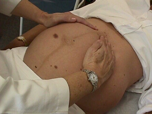 Fetal Orientation
The presentation (head first, breech first, transverse lie) and position
(anterior, posterior, transverse) can be determined in several ways:
- An ultrasound scan will confirm the presentation and position any time it is needed.
- An x-ray of the abdomen can provide nearly as much information as the ultrasound scan, but exposes both the mother and fetus to radiation and thus is rarely used.
- Clinical examination of the abdomen (Leopold's Maneuvers) can provide very reliable information, although the more experienced the examiner, the more reliable the information. Patient habitus also makes this exam easier or more difficult.